Module 01. Biomechanics
Decoupling biology from mechanics
A shift toward non-compressive biomechanical anchorage. A biological hypothesis grounded in vascular physics, bone histology, and two independent orthopedic precedents.
Conventional torque
>35–50 N·cm
Radial compression that exceeds the physiological capillary perfusion threshold.
RAMAX® insertion
<20 N·cm
Design parameter: low torque, aimed at limiting radial interference at the interface.
Vascular threshold
20–30 mmHg
Physiological limit of capillary collapse in non-expandable bone canals.
Active fixation
6 DOF
Architecture designed to restrain 6 degrees of freedom through a through-going transverse screw.
Scientific rationale
The physical principle in detail
The argument does not rest on an isolated claim. The underlying physics is compartment syndrome: any vascular bed confined within a rigid bony space collapses at pressures as low as 20-30 mmHg, regardless of the direction the compression comes from. In 1988, Perren et al. confirmed that this principle holds true in real bone: bone fixation by external compression induced measurable cortical necrosis, not a simple stress-protection response (Clin Orthop Relat Res 232:139–151) — the first direct proof that compressing cortical bone, in practice, does compromise its vascularization. The finding was independently confirmed by Uhthoff et al. in 1994 (J Bone Joint Surg 76-A:1507–1512). That same year, 1988, Brumback et al. consolidated a different engineering solution to the same problem: the statically locked intramedullary nail (J Bone Joint Surg 70:1453–1462), which anchors via a transverse canal crossed by a screw against the opposite cortex, without relying on sustained compression. RAMAX® applies that same physical principle — already demonstrated in real bone, and already solved with geometric anchorage in orthopedics for nearly four decades — to oral implantology; the GLP study with AnaPath does not aim to prove the general principle, but rather its translation to the scale of a dental implant.
Comparison
Conventional paradigm versus the RAMAX® System
Conventional paradigm — friction-based press-fit
Cortical ischemia
Radial compression from a tapered implant exceeds the capillary perfusion threshold, creating a risk of ischemia in the surrounding Haversian canals.
Microdamage and remodeling
Excessive torque can cause structural microdamage, forcing a prolonged osteoclastic resorption cycle before repair.
Stability dip (weeks 2–4)
A well-documented period of declining primary stability, before secondary biological stability matures.
Crestal bone loss
Resorption of a compromised cortical rim can expose the implant to the peri-implant sulcus (Chen et al., Periodontology 2000, 2026).
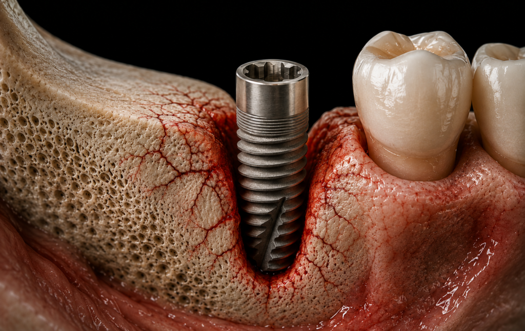
Simulation — vascular damage and microfractures from sustained radial compression.
RAMAX® System — design objective, pending validation
Low-torque insertion (<20 N·cm), designed to limit radial interference
The cylindrical body seats into the osteotomy without significant radial expansion force.
Predicted biological response
A reduction in the affected tissue area is predicted, to be confirmed through histological analysis.
Stability curve
An attenuated stability dip is postulated, subject to quantification in the preclinical study.
Crestal level
The absence of sustained compression — though not yet confirmed with our own data — is expected to contribute to long-term preservation of marginal bone.
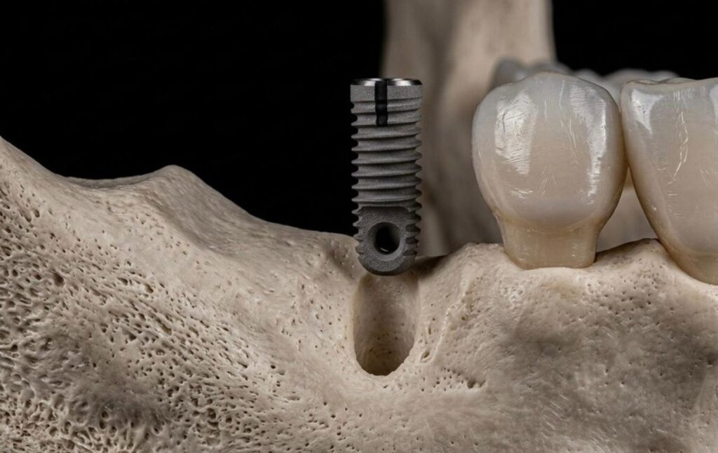
Implant body with apical transverse channel — passive insertion without radial expansion.
Stability dynamics
Hypothesis of biomechanical behavior
Theoretical model comparing the stability curve of friction-based systems (with a decline documented in the literature) against the curve postulated for RAMAX® — attenuated and more continuous, by decoupling primary fixation from the tissue undergoing remodeling.
Lateral biomechanical perspective: the transverse screw seeks primary stability through bicortical anchorage in the basal cortical bone, with the aim of decoupling fixation from crestal bone quality.
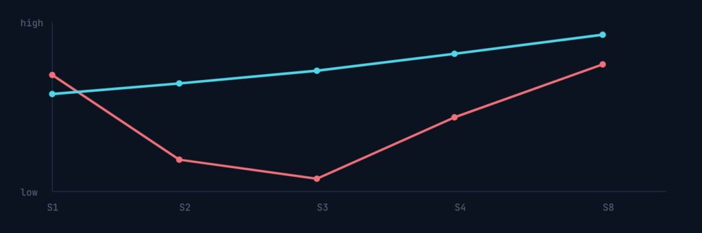
Chart note: theoretical model. Hypothesis subject to histomorphometric quantification in the GLP preclinical study with AnaPath; measured results are not yet available.
AnaPath Services GmbH
Real mandible model
Surgical sequence
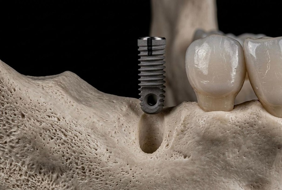
IMPLANT INSERTION — Passive placement in the recipient bed
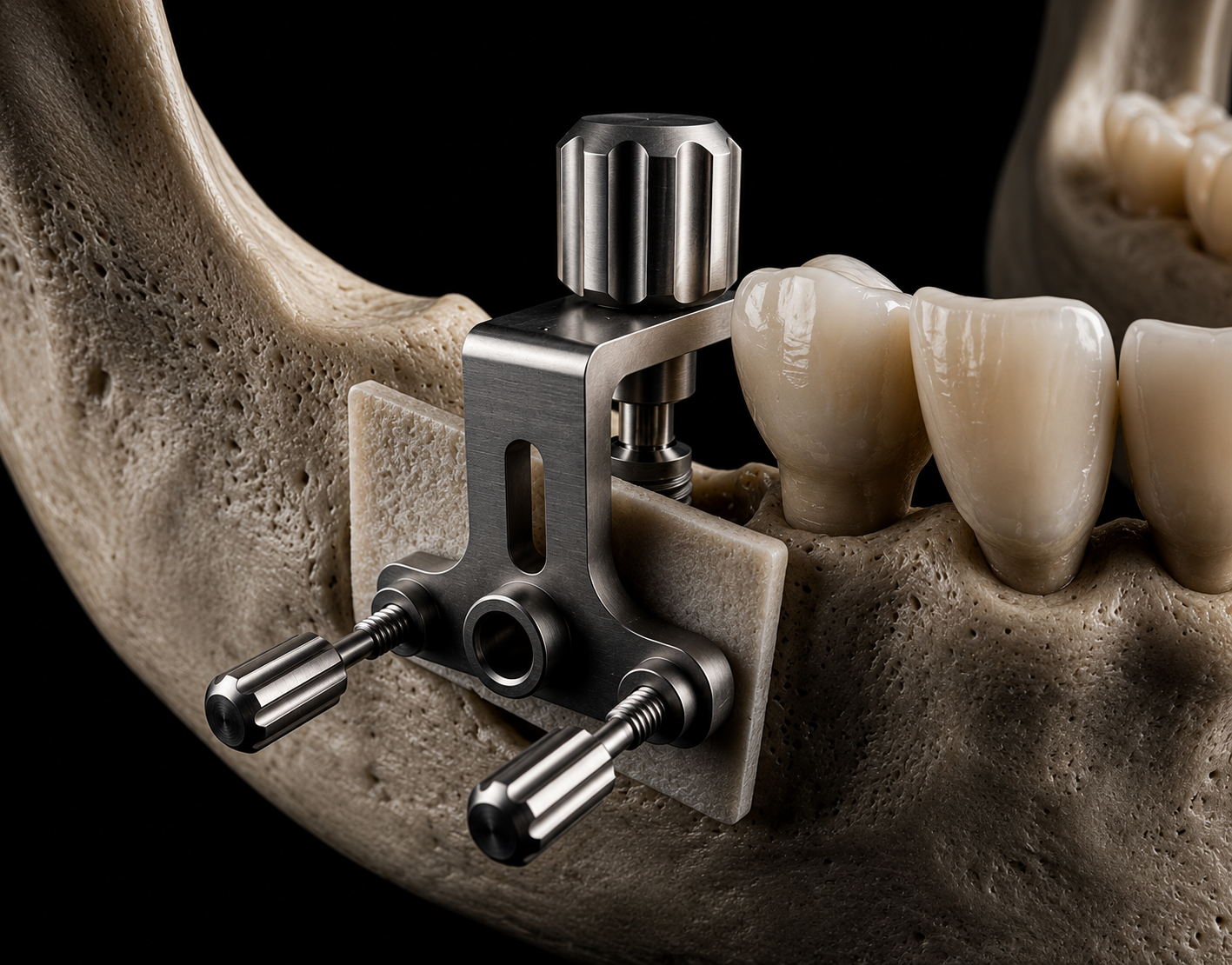
DRILLING GUIDE — With allogeneic cortical plate
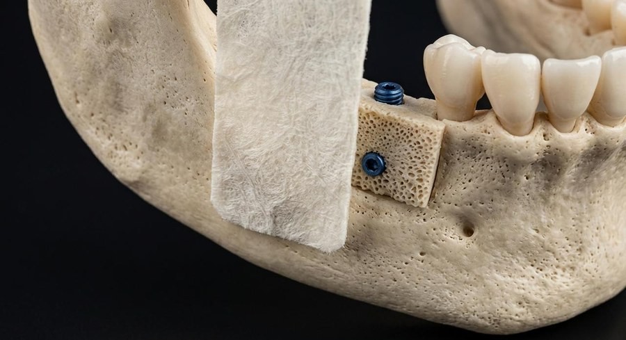
COLLAGEN MEMBRANE — With secondary screw
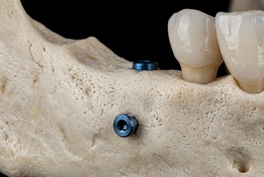
TARGET BIOLOGICAL OUTCOME